Histological Services
Tissue characterization procedures may include histological or immunohistochemical analysis to enrich the pathological donor data. We apply optimized protocols to visualize common hallmarks of disease such as βA4, α-syn, TDP43 and offer to validate and apply customized protocols for staining of specific targets of interest in fixed or freshly frozen tissue. In addition, other quantitative methods for expression of RNA or proteins are offered to explore the expression of potential therapeutic targets. Together this will facilitate optimal selection and characterization of tissue, at the level of the donor or tissue block.
Immunohistochemistry & immunofluorescence
- FFPE & cryo tissue block characterization
- Pathological screening of tissue blocks: aβ, α-synuclein, Phospho-Tau (AT8)
- Lesion characterization: human leukocyte antigen (HLA)/ proteolipid protein (PLP)
- Additional targets on request
- Immunostaining protocol development and validation
- Target validation/visualization
- Imaging with multispectral slide scanner and high-resolution fluorescent microscopy
- Image analysis
-01.png)
Example histological and immunohistochemical staining for neuropathological assessment of donors
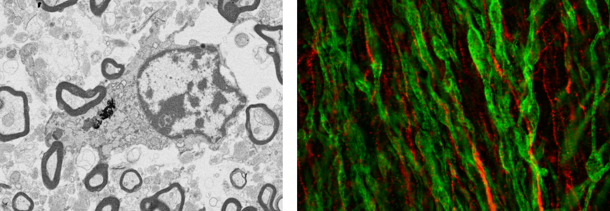
Advanced imaging procedures applied on NBB postmortem tissue. Left: Electron microscopy image on glutaraldehyde fixed tissue showing oligodendrocyte and axons in cross-section. The eletron microscopy image was generated by Dr. Wiebke Möbius, Max Planck Institute, Göttingen. Right: Stimulated emission depletion (STED) image optic nerve tissue in MS donor, visualizing Myelin Binding Protein (green) and Anti-Neurofilament antibody [SMI-312] (red); Image generated by Aletta van den Bosch, Netherlands Institute for Neuroscience.