Autopsy and dissection procedures
Since 1985, the NBB has obtained brain material from over 5000 donors. Due to the 24/7 availability of the NBB and VUmc staff, combined with the small size of the Netherlands, rapid autopsies with short post-mortem delays of three to ten hours are possible.
Once the brain has been removed it is macroscopically examined and dissected according to the clinical diagnosis and requests of researchers. On average some 90 tissue samples are frozen in liquid nitrogen or fixed in formalin. Before the brain is removed, CSF is extracted from the lateral ventricles with an 18GA 3.50 inch spinal needle and the pH of the CSF is measured. In addition, plasma and sometimes spinal cord and dorsal root ganglia are collected.

The neuropathologist
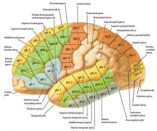
Then the external structures such as basal arteries, the pituitary gland and the olfactory bulb are removed. Next, the medulla oblongata is sampled, together with the cervical spinal cord and samples from the cerebellum. Then the brainstem is cut in half. The right side is put into 4% formaldehyde and from the left side the substantia nigra, locus coeruleus and dorsal vagus nucleus are rapidly frozen and stored. The cerebrum is cut in half and the right side is fixed in 4% formaldehyde. From the left side the cortex is sampled first, then the hippocampus and amygdala are dissected, after which the hypothalamus, thalamus and the basal nuclei are snap frozen. Finally, white and grey matter are harvested for tissue cultures. Brain regions are dissected according to the scheme below.
 |
Tissue treatment in case of multiple sclerosis
The clinical diagnosis multiple sclerosis (MS) requires a different protocol.
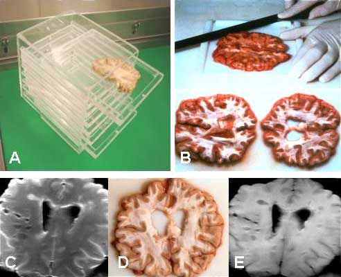
The brain is cut into coronal slices of 1 cm using a ‘brain slice holder’. In cooperation with the Department of Radiology of the VUmc, some of these slices are scanned using magnetic resonance imaging (MRI) to identify and dissect macroscopically invisible MS lesions. These lesions and some of the macroscopically visible lesions are split. One half is frozen and the other half is fixed in formalin (‘mirror blocks’). The remaining macroscopically visible lesions are frozen as a whole. The tissue in formalin is dissected after four weeks of fixation into a standard number of approximately eighteen regions for diagnostic purposes.
HomeBrain tissueDissection