Characterization of MS lesions
It is crucial that all MS tissue blocks are carefully characterized and the MS lesions, if present, are precisely characterized/staged 1–6 for white and grey matter lesions, including subscores for myeloid activity (see figure and table below).
This implies that sections of the formalin part of the mirror blocks (see more about mirror blocks below) containing MS lesions and NAWM dissected by MRI guidance, as well as all diagnostic mirror blocks from the brain stem, hippocampus, basal ganglia and spinal cord, are double-stained for PLP/HLA and Kluver (find an overview of all performed stainings here).
Sections with the HLA/PLP double staining are photographed and the characterization is indicated in codes of these photographs (figure and table below). This facilitates the search of MS tissue to address all different research questions (see publications).
In addition to the characterization of all MS tissue blocks, the final diagnosis of the donor will be determined by our neuropathologist based on the diagnostic blocks and 5 representative MS lesions of the patient, together with the clinical information that is summarized and translated into English by a clinical summarizer of the NBB.
 |
Figure 1: Classification of MS lesion and myeloid activity (scores 0. 0.5, 1 represent myeloid sub-scores 1, 2 and 3 in the official characterization) in the MS lesions according to Luchetti et al., 2018.
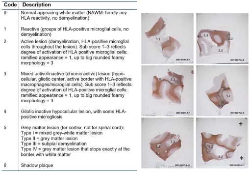 |
Table 1: Staging of MS lesions according to Luchetti et al., 2018. Lesion scores (1–6) and myeloid activation sub- scores (1–3 see figure 1) are indicated in photographs of HLA-PLP stained sections and used to select tissue for MS tissue applicants.
Mirror blocks
Lesions are detected in 1 cm coronal sections and are dissected. Lesions are detected either macroscopically or using MRI guidance, read more about this here. These dissected lesions are then divided in two blocks of 0.5 cm. These are mirror blocks: 0.5 cm is frozen in liquid nitrogen and 0.5 cm is fixed in formalin. Lesion characterization is performed on the formalin-fixed block, on sections cut from the side where the formalin fixed block bordered the frozen block. Nevertheless, please note that it is possible that the part of the lesion captured in the formalin fixed block, is not the exact same lesion type as in the frozen block. Therefor the NBB cannot guarantee that the frozen lesion blocks always contain the exact lesion type that was characterized based on the formalin fixed block.