Research output
Researchers about the importance of NBB-Psy
The data and tissue generated by NBB-Psy are crucial to allow advances in psychiatric research. Three researchers illustrate the importance of NBB-Psy below.
|
|
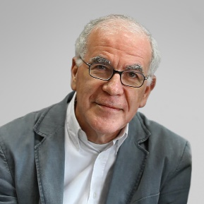
Dr. Karel Scheepstra – Psychiatrist and postdoctoral researcher“Disturbances in functioning in someone with depression can be so severe, that we should be able to find alterations in the brain.” |
|
|
Can you explain the importance of your research in some more detail? I am a psychiatrist and postdoctoral researcher working at the mood disorder clinic of the Amsterdam UMC (AMC location) and the Netherlands Institute for Neuroscience (NIN), Neuroimmunology group. To conduct sustainable research in the field of neurobiology of psychiatric disorders, a collection of excellent brain tissue is essential. Most of the current fundamental stress and depression research is done in vitro or in animal models, such as mice. However, the best model for human brain diseases is the human brain itself. An intricate and complex disease such as depression cannot be entirely simulated in the lab. One of my goals at the Netherlands Brain Bank (NBB) is selecting well-phenotyped donors that are suitable for research, as major depressive disorder is a heterogeneous disease with many comorbidities. Furthermore I research postmortem brain tissue of patients with a severe mood disorder. My interest is in biological psychiatry and my goal is to understand the ´severely depressed brain´. Someone with severe depression can be so ´paralyzed´, that even simple and vital actions such as sleep, food intake, self-care and even wanting to be alive, are not possible anymore. In my opinion, these disturbances in functioning can be so severe, that we should be able to find alterations in the brain. The severe and refractory depressions I treat in the clinic often need biological treatments, such as medication, electroconvulsive therapy and even deep brain stimulation. Globally, thousands of people die yearly of depression and the burden of disease is high, for both patients and their families. Insights in novel therapeutics and disease pathophysiology are urgently needed, as treatment resistance is common and occurs in up to 30% of depressed patients. Why did you choose to carry out your research at the Netherlands Brain Bank? The NBB is unique. The tissue is of high quality and there is extensive clinical information on the course of illness. Some donors were selected from cohorts and have undergone additional tests when alive, such as neuropsychological tests or neuroimaging. Since 2012, the Netherlands Brain Bank for Psychiatric disorders (NBB-Psy) has made substantial progress and many new donors were registered. The phenomenology of psychiatric disorders is extraordinarily heterogeneous and complex. Therefore I think it is important to have a clinician from the field of mental health working with the NBB, to bridge the gap between the psychiatric clinic and researchers. Years of fundamental research on the neurobiology of depression have given inconsistent and conflicting results. One of the reasons is the heterogeneity of the disease, lack of mental health knowledge in fundamental researchers and the quality of tissue and donors. My hope, however, is that in the coming decade we will be able to gain more insight into the pathophysiology of depression, using our growing collection of brain tissue combined with new molecular techniques. Also new types of promising biological treatments, such as transcranial magnetic stimulation or esketamine (a N-methyl-D-aspartate receptor antagonist), may give a new window into the neurobiology of depression. |
||

|
|
Lot de Witte - Assistant Professor, Icahn School of Medicine at Mount Sinai, New York City, United States of America“NBB-Psy enabled us to carry out research into changes in the brain’s immune system of patients suffering from schizophrenia, bipolar disorder or depression. The most abundant immune cells, microglia, did not show infection-like activation but changes related their communication with neurons.” |
|
|
Changes in the immune system in the brains of patients with a psychiatric disorder In contrast to brain disorders such as Multiple Sclerosis and Alzheimer’s Disease, we know very little about what goes wrong in the brains of patients with a psychiatric disorder. The past 10 years there has been an increasing interest in the potential role of the immune system. A reason for this interest is the increased incidence of immune system diseases in patients with psychiatric disorders. In addition, an association has been found between several psychiatric disorders and genes known for their important immune function. Therefore we started in 2014 with various projects to map the role of the immune system in psychiatric disorders. In this process the collaboration with the Netherlands Brain Bank (NBB) was crucial. Due to the efforts of the NBB and a large group of Dutch psychiatrists the number of brain donors with a psychiatric disorder has increased significantly within the NBB-Psy framework. This enabled us to carry out research into changes in the immune system of patients suffering from schizophrenia, bipolar disorder or depression. When we started the research the common hypothesis was that in the brains of people with a psychiatric disorder a mild form of inflammation may exist. Therefore various clinical studies with anti-inflammatory medication were being conducted. We looked predominantly at the microglia cells, the most abundant immune cell in the brain. These cells are not only involved in regulating an infection but also in neuronal development and function. We use frozen or fixed tissue and isolate microglia from the brain tissue so that we can study in detail what they look like and how they respond to their environment. Our findings [1] contradicted the leading hypothesis; the microglia of patients with a psychiatric disorder were not activated as one would expect during an infection but showed changes related to their communication with neurons. In our further research we explore how this affects the neurons and other brain cells. In addition, we use reprogramming protocols allowing us to generate microglia from stem cells. For this research we hope to use the induced pluripotent stem cells (iPSCs) generated within the NBB-Psy program. |
||

|
|
Dick F. Swaab, MD, PhD - Emer. Prof. of Neurobiology, Univ. of Amsterdam | Qiu Shi Professor of Zhejiang University, Hangzhou, P.R. China | Head Laboratory for Neuropsychiatric Disorders, Netherlands Institute for Neuroscience, Amsterdamalso emphasizes that careful matching and stratification of control and patient groups is paramount. For example in case of suicidal behavior one should control for the underlying psychiatric disorder. ‘In this way postmortem brain material …. will be a very important step towards a better understanding of the molecular changes at play both in suicide and in the underlying psychiatric disorder and may hopefully lead to novel, effective treatment strategies for the prevention of suicide.” |
|
|
Postmortem studies on suicide: complex but urgently needed Suicide is a major mental health problem. Yearly one million people die of suicide worldwide. Suicide medications lack efficacy, so molecular research on postmortem brain is urgently needed to find novel targets for preventive treatment. However, when we analysed the international literature on molecular alterations in the post-mortem brain of persons after accomplished suicide, the results appeared to differ strongly between different studies, often even in opposite directions, in spite of the fact that the patient groups had been carefully matched for factors like age, gender and post-mortem delay. Suicide is for the major part occurring in people that have underlying psychiatric disorders, from which major depression is the major one but certainly not the only disorder. We found that at least part of the confusion in the literature resulted from the fact that suicide has always been considered to be a symptom, and thus an integral part of major depression, so that in studies on depression, the group of depressed patients contained often both, major depression patients with and without suicide. As a result, post-mortem studies that claimed to have determined molecular alterations in relation to major depression, had in fact often selected depressed patients that had died all, or for a major part, from suicide. They were commonly compared to control subjects without any psychiatric disorder, thereby disregarding the fact that suicide per se may have influenced gene expression in their postmortem brains as well, and may thus have confounded earlier findings that were interpreted as being specific for depression alone. Secondly, studies that claimed to have shown changes in relation to suicide, had generally compared cases who following accomplished suicide to matched controls without any psychiatric disorder, thereby disregarding the fact that suicide may occur in different psychiatric disorders and conditions, including mood and anxiety disorders, but also e.g. in schizophrenia, personality- or substance abuse disorder. To date, very few papers have attempted to study neurobiological differences between suicide and the underlying psychiatric disorder. We have therefore proposed that in future postmortem studies e.g. on mood disorders, ideally, at least four groups should be distinguished: 1) age-matched controls without a brain disorder, 2) depressed patients who did not commit suicide and did not have suicidal ideations or attempts, and died from natural causes, 3) depressed patients with suicide attempts or ideations, but who died from natural causes, and 4) depressed patients with accomplished suicide. While brain material for such studies may not be easy to collect, it will be a very important step towards a better understanding of the molecular changes at play both, in suicide and in the underlying psychiatric disorder and may hopefully lead to novel, effective treatment strategies for the prevention of suicide. Zhao J, Lucassen PJ, Swaab DF. Suicide Is a Confounder in Postmortem Studies on Depression. Biol Psychiatry. 2019 Nov 15;86(10):e37-e40. doi: 10.1016/j.biopsych.2019.04.015. Epub 2019 Jun 18. PMID: 31227102. |
||
Different applications NBB-Psy tissue
The below publications illustrate the use of NBB brain tissue for different applications such as identifying disease and drug targets and pathways as well as biomarker validation.
Brain tissue can be used to identify molecular targets and pathways. DNA methylation of 4 regions was differentially regulated in the gyrus frontalis medialis (GFM) and gyrus temporalis superior (GTS) grey and white matter of schizophrenic compared to control donors. These regions were within or close to the genes KLF9, SPRED2 (brain cell development, migration and differentiation) and SFXN1 (mitochondrial function), which were also differentially regulated in the different brain tissues of SCZ patients. These results indicate the role for these genes and DNA methylation in the pathophysiology of SCZ (Berdenis van Berlekom et al., 2021).
A study illustrating the use of postmortem brain tissue for biomarker identification showed that consistency of protein expression could be validated with brain tissue of MDD patients and controls. Plasma (inter-alpha-trypsin inhibitor heavy chain 4) ITIH4, a neuroinflammation marker, and vitamin D-binding protein (VDB) levels, affecting vitamin D levels and inflammation, were identified in psychiatric patients using protein profiling and shown to be specific for MDD. These two proteins were also significantly increased in the fresh frozen dorsolateral prefrontal cortex (DLPFC) of NBB donors with MDD compared to controls and protein levels correlated with neuropsychological assessments (Shi et al., 2020).
Brain tissue can also be used to elucidate the brain pathway and efficacy of medication, such as the antipsychotic drug clozapine. Using NBB tissue, one study confirmed the strong effect of clozapine on retinoic acid (RA) catabolism as compared to other psychotropic drugs (Regen et al., 2020).
Publications
A few key publications made possible by the NBB-Psy program are listed below. A yearly list of all publications, including those using NBB-Psy tissue can be found on the NBB’s publication page.
- Berdenis van Berlekom, A., Notman, N., Sneeboer, M.A., Snijders, G.J., Houtepen, L.C., Nispeling, D.M., He, Y., Dracheva, S., Hol, E.M., Kahn, R.S., de Witte, L.D., Boks, M.P., Psychiatric Donor Program of the Netherlands Brain Bank (NBB-PSY), 2021. DNA methylation differences in cortical grey and white matter in schizophrenia. Epigenomics 13, 1157–1169. https://doi.org/10.2217/epi-2021-0077
- Böttcher, C., Schlickeiser, S., Sneeboer, M. A. M., Kunkel, D., Knop, A., Paza, E., Fidzinski, P., Kraus, L., Snijders, G. J. L., Kahn, R. S., Schulz, A. R., Mei, H. E., Netherlands Brain Bank for Psychiatry, Hol, E. M., Siegmund, B., Glauben, R., Spruth, E. J., de Witte, L. D., & Priller, J. (2019). Human microglia regional heterogeneity and phenotypes determined by multiplexed single-cell mass cytometry. Nature Neuroscience, 22(1), 78–90. https://doi.org/10.1038/s41593-018-0290-2
- Sneeboer, M. A. M., Snijders, G. J. L. J., Berdowski, W. M., Fernández-Andreu, A., Psychiatric Donor Program of the Netherlands Brain Bank (NBB-Psy), Mierlo, H. C. van, Berlekom, A. B. van, Litjens, M., Kahn, R. S., Hol, E. M., & Witte, L. D. de. (2019). Microglia in post-mortem brain tissue of patients with bipolar disorder are not immune activated. Translational Psychiatry, 9. https://doi.org/10.1038/s41398-019-0490-x
- Sneeboer, M. A. M., van der Doef, T., Litjens, M., Netherlands Brain Bank for Psychiatry, Melief, J., Hol, E. M., Kahn, R. S., & de Witte, L. D. (2020). Microglial activation in schizophrenia: Is translocator 18 kDa protein (TSPO) the right marker? Schizophrenia Research, 215, 167–172. https://doi.org/10.1016/j.schres.2019.10.045
- Snijders, G. J. L. J., Sneeboer, M. A. M., Fernández-Andreu, A., Udine, E., Psychiatric donor program of the Netherlands Brain Bank (NBB-Psy), Boks, M. P., Ormel, P. R., van Berlekom, A. B., van Mierlo, H. C., Bӧttcher, C., Priller, J., Raj, T., Hol, E. M., Kahn, R. S., & de Witte, L. D. (2020). Distinct noninflammatory signature of microglia in post-mortem brain tissue of patients with major depressive disorder. Molecular Psychiatry. https://doi.org/10.1038/s41380-020-00896-z